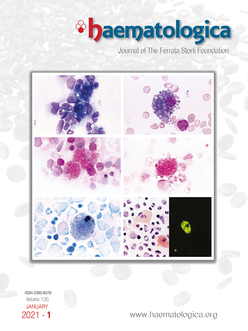
Sea-blue histiocytosis, also called syndrome of the sea-blue histiocyte, is a very rare lysosomal storage disease, characterized by distinctive morphological features of storage cells in the bone marrow (Figure), liver, spleen, and other organs, due to the accumulation of ceroid or lipofuscin in their cytoplasm. Bone marrow sea-blue histiocytes are filled with granules that stain blue or blue-green with May-Grünwald Giemsa stain (Figure A, B) or other panoptic stains, while they are brown in unstained films. Histiocyte granules are generally periodic acid Schiff positive (Figure C) and are sometimes iron positive. The histiocyte granules stain with Ziehl-Nielsen (acid fast) stain (Figure D), with phospholipids stained with Nile blue sulphate stain (Figure E) and lipids stained with Oil red O stain (Figure F, left). Moreover, under a fluorescence microscope, histiocytes show strong autofluorescence (Figure F, right).
Figure.
Footnotes
Correspondence
References
- Invernizzi R. Storage diseases. Haematologica. 2020; 105(Suppl 1):S255-260. https://doi.org/10.3324/haematol.2019.232124PubMedPubMed CentralGoogle Scholar
Data Supplements
Figures & Tables
Article Information

This work is licensed under a Creative Commons Attribution-NonCommercial 4.0 International License.

